
The Immunological Basis of Allergic Persistence and Resolution: A Comprehensive Analysis of Postnatal Adaptation, Cellular Ontogeny, and the Maintenance of Pathogenic Memory
Table of Contents
- Executive Summary
- Chapter 1: The Ontogeny of Immunity and the Neonatal Window
- Chapter 2: The Players – Origins, Education, and Cellular Architecture
- Chapter 3: The Mechanism of Memory – The Journey to Persistence
- Chapter 4: The Role of T Cells in the Bone Marrow – The “Nurse Cell” Function
- Chapter 5: Immunotherapy (AIT) – Rewiring the System
- Conclusion
- Summary Table: The Cellular “Dance” in Allergy
- References
Executive Summary
The human immune system is a sophisticated biological recognition engine, evolved to navigate a complex landscape of self-antigens, harmless environmental proteins, and pathogenic threats. In health, this system maintains a delicate equilibrium, deploying aggressive inflammatory responses against invaders whilst preserving tolerance towards benign substances. Allergic disease represents a catastrophic failure of this decision matrix, a disorder of “mistaken identity” in which innocuous proteins, such as pollen or peanut antigens, trigger a violent, protective Th2-type immune response originally evolved to defend against helminthic parasites.
This report provides an exhaustive examination of the immunological mechanisms underpinning allergy, tracing the trajectory from the neonatal period to the establishment of lifelong pathogenic memory. We dissect the critical “Th2-to-Th1 switch” that occurs post-birth, and the specific failures in dendritic cell maturation and cytokine signalling that arrest the immune system in an atopic state. We explore the ontogeny of the key cellular players, T cells and B cells, mapping their education in primary lymphoid organs and their corruption in the allergic cascade.
A central focus of this analysis is the “mechanism of memory.” We challenge traditional dogmas about the residency of IgE-producing cells, integrating emerging evidence suggesting that the spleen, rather than the bone marrow, may serve as the primary reservoir for mature, long-lived IgE plasma cells. Furthermore, we elucidate the complex “nurse cell” functions within survival niches, revealing how regulatory T cells (Tregs) and eosinophils, typically viewed through the lens of inflammation or suppression, act as guardians that paradoxically sustain pathogenic antibody production. Finally, we evaluate the molecular mechanisms of Allergen Immunotherapy (AIT), analysing its capacity to rewire this entrenched system through metabolic reprogramming, isotype switching, and the potential therapeutic depletion of the long-lived plasma cell reservoir.
Chapter 1: The Ontogeny of Immunity and the Neonatal Window
The genesis of allergic disease is often traced to the earliest moments of extrauterine life. The neonatal immune system is not merely an immature version of the adult system but a specialised, distinct state programmed for the unique requirements of gestation. Understanding the transition from this foetal state to a competent adult phenotype is essential for grasping the pathophysiology of allergy.
1.1 The Foetal Immune State: A Necessity of Tolerance
During pregnancy, the maternal immune system faces a formidable biological paradox: it must tolerate the foetus, a semi-allograft expressing paternal alloantigens, whilst retaining the capacity to fight infection. To prevent rejection, the maternal-foetal interface is maintained in a state of profound immunosuppression, specifically dominated by a Th2-biased cytokine environment.
Th1 cytokines, such as Interferon-gamma (IFN-γ) and Tumour Necrosis Factor-alpha (TNF-α), are potently inflammatory and cytotoxic. Their unrestricted expression at the decidual interface could damage the placental trophoblast and induce spontaneous abortion. Consequently, the foetal immune system develops under a “default” Th2 setting.1 This is not a passive absence of Th1 immunity but an active, epigenetically reinforced programme.
Epigenetic Poising:
Analysis of neonatal CD4+ T cells reveals a distinct chromatin landscape. The Th2 cytokine locus, which encodes Interleukin-4 (IL-4), IL-5, and IL-13, is epigenetically “poised” for transcription. Specifically, the Conserved Non-Coding Sequence-1 (CNS-1) region within the Th2 locus is hypomethylated (open) in neonatal T cells.1 In contrast, the promoter regions for IFN-γ are often hypermethylated (closed).4 This configuration ensures that if the foetus encounters an antigen in utero, the immune system defaults to a non-damaging Th2 response, preserving the pregnancy at the cost of potential future atopy.
1.2 The Postnatal Switch: The “Training Signal”
Upon delivery, the neonate moves from a sterile, antigen-restricted environment into a world teeming with microbial life. This exposure acts as the critical “training signal” required to rebalance the immune system. The transition from the foetal Th2 bias to a balanced Th1/Th2/Treg profile is dependent on specific innate immune signals, primarily driven by dendritic cell (DC) maturation.
The CD8α+ Dendritic Cell Delay:
A key mechanistic bottleneck in this transition is the functional immaturity of neonatal dendritic cells. Specifically, the CD8α+ CD4– subset of DCs, which is the primary physiological source of Interleukin-12 (IL-12), is quantitatively and functionally limited at birth.1 IL-12 is the master cytokine required to drive naïve T cells towards a Th1 phenotype and, crucially, to stabilise that phenotype by suppressing Th2 signalling machinery.
The Heteroreceptor Apoptosis Mechanism:
The failure to produce sufficient IL-12 in the immediate neonatal period has profound consequences for T cell survival. In the absence of IL-12, neonatal Th1 cells fail to downregulate the expression of the IL-13 receptor alpha-1 chain (IL-13Rα1).1 This receptor subunit pairs with the ubiquitously expressed IL-4 receptor alpha (IL-4Rα) to form a high-affinity heteroreceptor (IL-4Rα/IL-13Rα1) on the surface of the developing Th1 cell.
This creates a molecular “kill switch”:
- Primary Exposure: The neonate encounters an antigen. Due to low IL-12, Th1 cells differentiate but retain the IL-13Rα1 heteroreceptor.
- The Th2 Response: Simultaneously, the epigenetically poised Th2 cells differentiate and prepare to secrete cytokines.
- Secondary Challenge: Upon re-exposure to the antigen, Th2 cells rapidly secrete IL-4.
- Apoptotic Deletion: This IL-4 binds to the heteroreceptor on the vulnerable Th1 cells. Instead of stimulating proliferation, this signal triggers apoptosis (programmed cell death).1
This mechanism effectively “prunes” the Th1 arm of the immune response upon secondary exposure, reinforcing the default Th2 bias. The “switch” to a healthy, non-allergic phenotype depends on the timely maturation of DCs and the accumulation of sufficient IL-12 to break this cycle by downregulating the heteroreceptor.1
1.3 The Hygiene Hypothesis and Microbial Metabolites
The “Hygiene Hypothesis” posits that a lack of early microbial exposure leads to a failure in this maturation process. Modern research has elucidated the molecular mechanism behind this, centring on the interaction between the gut microbiome and the epigenetic regulation of T cells. Recent advances in immunology research continue to shed light on these complex interactions.
Short-Chain Fatty Acids (SCFAs) as Epigenetic Modulators:
Commensal bacteria, particularly those of the Clostridia class and Bacteroidetes phylum in the gut, ferment indigestible dietary fibres into Short-Chain Fatty Acids (SCFAs) such as butyrate, propionate, and acetate.5 These metabolites are not merely energy sources for colonocytes but potent signalling molecules that influence immune tolerance.
HDAC Inhibition and FoxP3 Induction:
Butyrate acts as a Histone Deacetylase (HDAC) inhibitor.6 HDACs are enzymes that remove acetyl groups from histone proteins, causing chromatin to condense and silencing gene expression. By inhibiting HDACs (specifically HDAC1 and HDAC3), butyrate maintains high levels of histone acetylation at the Foxp3 gene locus.
- FoxP3: This transcription factor is the master regulator of Regulatory T cells (Tregs).
- Mechanism: Increased acetylation at the Foxp3 promoter and conserved non-coding DNA elements (CNS) render the chromatin accessible for transcription factors. This promotes the differentiation of naïve T cells into peripheral Tregs (pTregs) and stabilises their suppressive phenotype.9
In infants with a compromised microbiome (e.g., due to Caesarean section, antibiotic use, or low-fibre diet), SCFA levels are reduced. This leads to increased HDAC activity, silencing of the Foxp3 locus, and a failure to generate the robust Treg population needed to control the default Th2 bias. Without this regulatory brake, the immune system remains “stuck” in the atopic mode, interpreting harmless environmental proteins as dangerous parasites.
Chapter 2: The Players – Origins, Education, and Cellular Architecture
To understand the pathology of allergy, one must first map the ontogeny of the cellular protagonists: the T cells and B cells. Their “education” in primary lymphoid organs determines whether they become protectors of the host or pathogenic agents of allergic disease.
2.1 T Cells: The Generals of the Immune Response
Origin: T cell progenitors originate in the bone marrow from Haematopoietic Stem Cells (HSCs) but must migrate to the thymus for maturation, a journey that is critical for the establishment of self-tolerance.11
Education in the Thymus:
The thymus is a stratified organ where T cell “schooling” occurs in two distinct phases, ensuring both competence and safety.
- Positive Selection (The Competence Test): Occurring in the thymic cortex, double-positive (CD4+CD8+) thymocytes interact with Cortical Thymic Epithelial Cells (cTECs) expressing MHC molecules. T cells must demonstrate the ability to bind to self-MHC with at least weak affinity. Those that bind successfully receive survival signals (primarily IL-7), whilst those that cannot bind die by “death by neglect”.11
- Negative Selection (The Safety Test): Surviving thymocytes migrate to the thymic medulla. Here, they encounter Medullary Thymic Epithelial Cells (mTECs) and dendritic cells presenting a vast array of “self” antigens. This display is facilitated by the transcription factor AIRE (Autoimmune Regulator), which enables mTECs to express tissue-restricted antigens (e.g., insulin and thyroid proteins) that are normally expressed only in peripheral organs.11 If a T cell receptor (TCR) binds too strongly to a self-antigen, the cell is deleted via apoptosis (clonal deletion) to prevent autoimmunity.
The Treg Deviation:
Importantly, some T cells that bind self-antigens with intermediate to high affinity are not deleted. Instead, they are diverted into the Natural Regulatory T cell (nTreg) lineage.13 These FoxP3+ cells exit the thymus specifically programmed to suppress immune responses against the self-antigens they recognised. In allergy, the failure often lies not in these nTregs, but in the insufficient generation of inducible Tregs (iTregs) in the periphery, which are needed to tolerate non-self environmental antigens (allergens).14
The Allergic General (Th2 Differentiation):
In the context of atopy, a naïve CD4+ T cell (Th0) in secondary lymphoid organs differentiates into a pathogenic Th2 effector. This process is driven by the cytokine IL-4 (often derived from innate lymphoid cells, basophils, or mast cells) and the transcription factor GATA3. These Th2 cells orchestrate the allergic response by secreting specific cytokines:
- IL-4: The primary driver of B cell class switching to IgE.
- IL-5: The major survival and activation factor for eosinophils.
- IL-13: A mediator of mucus hypersecretion, airway hyperresponsiveness, and tissue remodelling.15
2.2 B Cells: The Factories
Origin: B cells develop entirely within the bone marrow. Their education involves rearranging immunoglobulin genes (V, D, and J segments) using the RAG1 and RAG2 enzymes to create a unique B Cell Receptor (BCR).
Central Tolerance: Like T cells, immature B cells in the marrow are tested for self-reactivity. Those binding self-antigens with high affinity undergo receptor editing (trying a new light chain gene rearrangement) or apoptosis.11
The IgE Class Switch:
Upon activation by a Th2 cell (via CD40L-CD40 interaction and IL-4/IL-13 signalling), a mature B cell in the lymph node undergoes Class Switch Recombination (CSR). It excises the gene segments for IgM and IgD and brings the variable region adjacent to the epsilon (ε) heavy chain constant region.16
Sequential vs. Direct Switching:
Recent research indicates two pathways for IgE generation:
- Direct Switching: IgM → IgE. This pathway is rapid but often yields low-affinity antibodies.
- Sequential Switching: IgM → IgG1 → IgE. Emerging evidence suggests that this is the primary pathway for generating high-affinity, pathogenic IgE in persistent allergy.16 The IgG1 intermediate phase allows the B cell to undergo extensive affinity maturation in the Germinal Centre (GC) before the final switch to IgE. This is crucial because IgE+ B cells themselves tend to exit the GC reaction early or undergo apoptosis, limiting their ability to mature affinity directly.16
Chapter 3: The Mechanism of Memory – The Journey to Persistence
The defining characteristic of chronic allergy is its persistence. An individual sensitised to peanuts or pollen may remain allergic for decades, even in the absence of constant exposure. This persistence is mediated by the establishment of immunological memory, specifically through Long-Lived Plasma Cells (LLPCs). The journey of an IgE B cell to this state differs significantly from other isotypes, a subject of intense recent investigation.
3.1 The Bifurcation of Fate: Memory B Cells vs. Plasma Cells
Upon sensitisation in secondary lymphoid organs (lymph nodes or spleen), activated B cells split into two distinct lineages with divergent destinies:
A. Memory B Cells (The Patrollers):
These cells return to a resting state and circulate in the blood and lymph. They express surface IgE (or often IgG1 in “covert” memory) but do not actively secrete antibodies. They are “patrollers” waiting for re-exposure to the allergen. Upon encountering the antigen again, they rapidly proliferate and differentiate into antibody-secreting plasmablasts. Recent single-cell transcriptomic studies suggest that true IgE+ memory B cells are extremely rare; instead, the “memory” of IgE responses is often held within an IgG1+ memory B cell compartment poised to switch to IgE upon reactivation.16
B. Plasma Cells (The Residents):
These are terminal effectors committed to massive antibody secretion (up to 10,000 molecules per second). They undergo significant morphological changes, expanding their endoplasmic reticulum to support protein synthesis. They downregulate surface BCR and upregulate the transcription factor Blimp-1 (Prdm1), which orchestrates the secretory machinery and silences the gene programmes associated with B cell identity (e.g., Pax5, Bcl6).20
3.2 The Controversy of the IgE Niche: Bone Marrow vs. Spleen
Standard immunological dogma holds that all long-lived plasma cells migrate to the bone marrow (BM), where specialised stromal cells provide survival signals (the “survival niche”) allowing them to live for years. This is well-established for IgG and IgA antibodies. However, for IgE, the picture is far more complex and controversial.
The Traditional View:
IgE plasma cells, like their IgG counterparts, migrate to the bone marrow and reside there as LLPCs. This reservoir explains the lifelong presence of serum IgE in atopic individuals.
The Emerging Evidence (The Spleen Hypothesis):
Recent “deep research” utilising timestamping models and single-cell RNA sequencing challenges the exclusivity of the bone marrow for IgE longevity. Studies by Limnander, Orengo, and others21 suggest a revised model:
- Short-Lived Nature: Most IgE plasma cells generated during a primary response are intrinsically short-lived and pro-apoptotic. They express high levels of surface BCR, and paradoxically, cross-linking of this receptor can induce apoptosis rather than activation.24
- Impaired Migration: IgE plasma cells often exhibit low expression of CXCR4, the chemokine receptor required for homing to the CXCL12-rich bone marrow niche. This renders them “unfit” for BM migration compared to IgG cells.21
- Splenic Residency: Advanced tracking reveals that a significant population of mature, long-lived IgE PCs resides in the spleen and lymph nodes rather than the bone marrow.21
- Transcriptional Profile: These stable IgE PCs exhibit a distinct phenotype: MHCIIlow CD93+ CD98high. This profile indicates high protein synthesis capacity (CD98 is an amino acid transporter) but a loss of antigen-presentation capability (MHCII low), marking them as fully differentiated factories.21
Implications for Persistence:
This finding implies that the “reservoir” of allergic memory may be more distributed than previously thought. The spleen, often viewed merely as a site of induction or filtration, acts as a major sanctuary for IgE production in persistent allergy. This may explain why bone marrow aspirates from allergic patients sometimes show lower IgE PC frequencies than expected relative to their serum IgE titres.20
3.3 Mechanisms of Longevity: The Survival Code
Whether in the bone marrow or the spleen, LLPCs cannot survive autonomously. They require an extrinsic “survival code” provided by the niche.
- Mcl-1 vs. Bcl-2 Dependency: IgE LLPCs appear to be hierarchically more reliant on the anti-apoptotic molecules Bcl-2 and Bcl-xL for their survival, whereas other plasma cell isotypes rely more heavily on Mcl-1.26 This differential dependency creates a potential therapeutic window: Bcl-2 inhibitors (such as navitoclax) could theoretically purge the allergic memory reservoir whilst sparing other protective antibodies.26
- The BCR Paradox: Unlike IgG PCs, which downregulate surface BCR almost completely, IgE PCs often retain significant surface BCR expression. Whilst this can serve as a “death receptor” if cross-linked by antigen, within the protected environment of the survival niche, these cells are shielded from such signals, allowing them to pump out IgE indefinitely.24
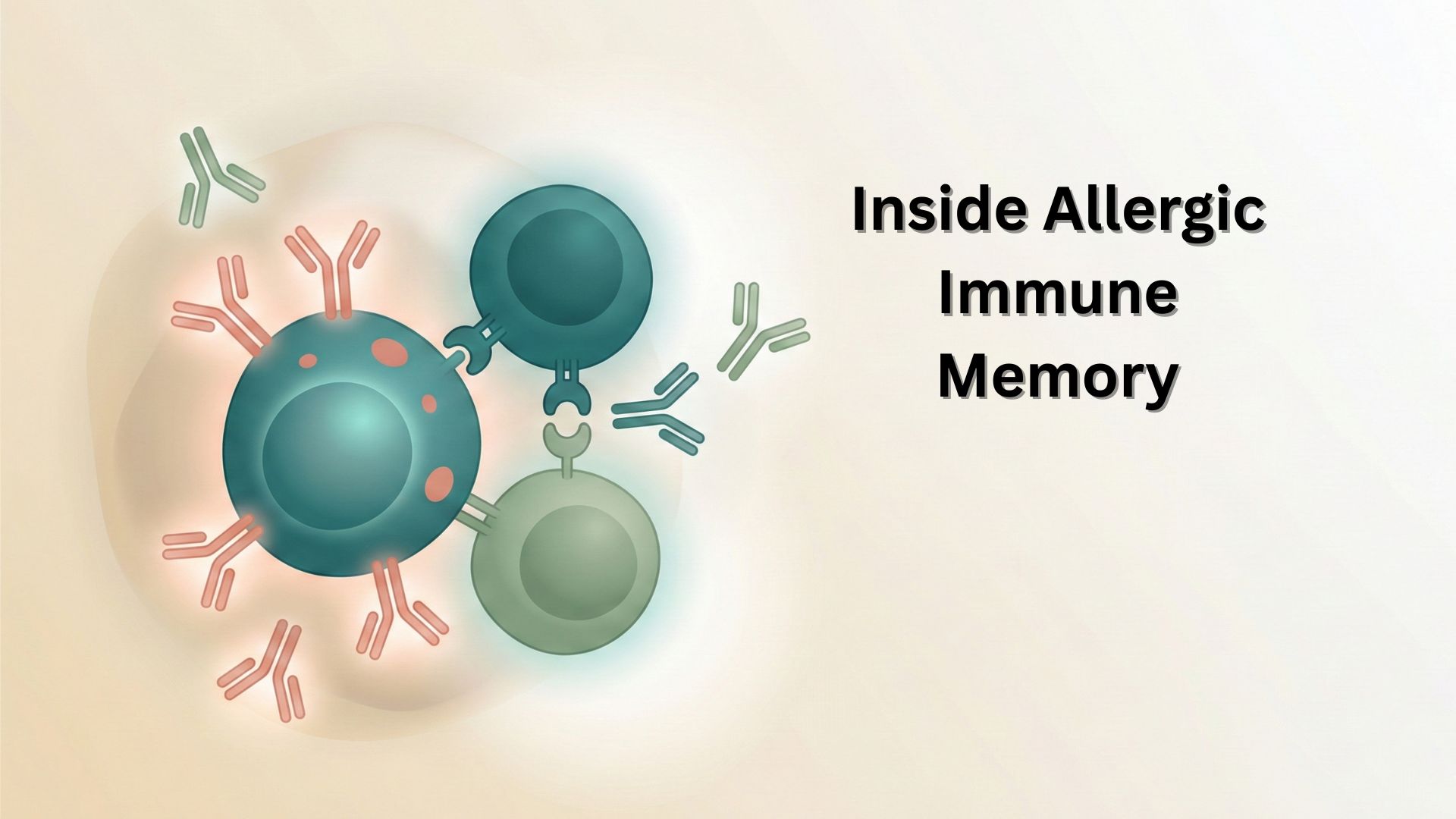
Chapter 4: The Role of T Cells in the Bone Marrow – The “Nurse Cell” Function
We specifically highlighted the movement of T cells to the bone marrow. This is a critical and sophisticated aspect of immunological memory. The bone marrow is not just a factory for creating cells; it is a sanctuary for preserving them.
4.1 The “Nurse Cell” Concept
The term “nurse cell” was originally applied to thymic epithelial cells encasing developing thymocytes. However, in the context of the plasma cell niche, it refers to the haematopoietic and stromal cells that physically nurture the LLPC and provide the signals necessary to inhibit apoptosis.
T Regulatory Cells (Tregs) as Guardians:
Research has identified a specialised population of Tregs residing in the bone marrow. Unlike their peripheral counterparts, which suppress inflammation, BM Tregs function as critical support cells for LLPCs.28 Understanding these mechanisms is crucial for developing new immunotherapy approaches.
- Co-localisation: Intravital imaging shows that BM Tregs cluster directly adjacent to plasma cells and CD11c+ dendritic cells.28
- Mechanism of Support: These Tregs secrete cytokines and provide contact-dependent signals (via CTLA-4) that modulate the local environment. Crucially, Treg depletion leads to a precipitous loss of BM plasma cells.29 They essentially maintain the “immune privilege” of the niche, preventing inflammatory damage and delivering survival signals.
- The “Double-Edged Sword”: In health, this mechanism protects vaccine-induced immunity (e.g., tetanus or measles antibodies). In allergy, these same Tregs may inadvertently protect pathogenic IgE LLPCs, acting as their “bodyguards” against turnover or therapeutic depletion.29
4.2 The Multicomponent Niche
The survival niche is a complex multicellular ecosystem. T cells are not the only guardians.
- Eosinophils: Historically, eosinophils were the first haematopoietic cells identified as obligate partners for LLPCs in the bone marrow. They secrete APRIL (A Proliferation-Inducing Ligand) and IL-6, which are essential survival factors for plasma cells.31 Since allergic inflammation is characterised by high levels of IL-5 and eosinophilia, allergic patients may possess an “enriched” or hypertrophic survival niche in the bone marrow. This abundance of eosinophils could further promote the retention and survival of IgE plasma cells, creating a self-reinforcing cycle of disease.
- Stromal Cells: CXCL12-producing reticular stromal cells form the physical scaffold of the niche. The receptor CXCR4 on the plasma cell serves as the “anchor” that holds the cell in place.33 The reduced expression of CXCR4 on some IgE plasma cells may explain their preferential retention in the spleen (which utilises different chemokine gradients) rather than the bone marrow.21
Chapter 5: Immunotherapy (AIT) – Rewiring the System
Allergen Immunotherapy (AIT), or desensitisation (allergy shots/drops), is currently the only disease-modifying treatment for allergy. It does not merely mask symptoms like antihistamines; it attempts to reverse the biological errors described in the previous chapters.
5.1 Mechanisms of Tolerance Induction
AIT works by flooding the system with antigen (e.g., pollen, venom) in a controlled, escalating manner. This high-dose exposure forces a deviation from the pathogenic Th2 response to a regulatory phenotype.
1. Treg Induction (The Master Switch):
High-dose antigen exposure drives the generation of Tr1 cells (Type 1 Regulatory T cells) and FoxP3+ Tregs.35 These cells secrete high levels of IL-10 and TGF-β.
- IL-10 Effect: It suppresses Th2 cytokine production, inhibits mast cell degranulation, and suppresses T cell proliferation. Crucially, it acts directly on B cells to alter their class-switching programme.
2. The IgG4 Blockade (The B Cell Switch):
IL-10 drives B cells to switch from producing IgE to producing IgG4.36
- IgG4 Function: IgG4 is a unique “blocking antibody.” It is non-inflammatory (it does not fix complement) and has high affinity for the allergen. Because IgG4 is present in serum at much higher concentrations than IgE after therapy, it intercepts the allergen before it can reach the IgE bound to mast cells. This prevents the cross-linking of FcεRI receptors and subsequent degranulation.
- The Ratio: Success in AIT is often measured by the increase in the IgG4:IgE ratio.
5.2 Does AIT Deplete the Bone Marrow Reservoir?
This question represents the frontier of allergy research and the “Holy Grail” of a permanent cure.
- The Limitation: Standard AIT is highly effective at inducing tolerance (suppression) and generating blocking antibodies. However, it rarely completely eliminates the reservoir of long-lived IgE plasma cells in the bone marrow or spleen. This explains why IgE titres often remain detectable even in patients who have been successfully treated, and why symptoms can return years after stopping therapy.27 The therapy functionally silences the disease mechanisms without fully deleting the pathogenic memory source.
The “Killer Treg” Hypothesis:
Emerging evidence suggests that specific subsets of induced Tregs (and regulatory B cells) may have cytotoxic capabilities. These cells can secrete Granzyme B and Perforin, molecules typically associated with Cytotoxic T Lymphocytes (CD8+), to directly kill effector B cells or plasma cells.40
- Mechanism: Granzyme B can enter the target cell and cleave caspases, inducing apoptosis.
- Therapeutic Potential: If AIT protocols could be optimised to specifically induce these “killer Tregs” or “killer Bregs,” it might be possible to actively purge the pathogenic IgE plasma cell niche, rather than just suppressing it.
5.3 Future Directions: Targeting the Niche
To achieve a permanent cure, future therapies must evict the LLPC from its survival niche. Ongoing research at leading immunology conferences continues to explore these innovative approaches.
- Navitoclax (Bcl-2 Inhibition): Since IgE LLPCs are uniquely reliant on Bcl-2 (unlike IgG cells, which use Mcl-1), Bcl-2 inhibitors could selectively purge the allergic memory reservoir whilst sparing protective immunity.26
- BCMAxCD3 Bispecific Antibodies: Adapted from multiple myeloma treatment, these engineered antibodies physically tether a T cell (via CD3) directly to a plasma cell (via BCMA), forcing the T cell to kill the plasma cell.18 In mouse models, this approach has successfully depleted IgE memory, offering hope for a “reset” of the allergic immune system.27
Conclusion
The transition from a healthy newborn immune system to a chronically allergic one is a multistep failure of adaptation. It begins with the inability to switch off the foetal Th2 programme due to insufficient microbial signals (IL-12/SCFA). It is consolidated by the “miseducation” of B cells, which switch to IgE and differentiate into Long-Lived Plasma Cells. These cells find sanctuary in survival niches within the spleen and bone marrow, protected by a complex support network of stromal cells, eosinophils, and “nurse” Tregs.
Understanding this cellular “dance” reveals why simple antihistamines fail to cure, they treat the downstream symptom, not the upstream source. True resolution of allergy requires not just blocking the mediator, but disrupting the survival niche itself, either through metabolic retraining (AIT) or targeted molecular deletion of the pathogenic memory reservoir.
Summary Table: The Cellular “Dance” in Allergy
| Stage | Location | What Happens? | Key Molecular Players |
|---|---|---|---|
| 1. Origin | Bone Marrow | B Cell and T Cell progenitors are born. VDJ recombination creates receptor diversity. | RAG1/2, TdT |
| 2. Education | Thymus | T Cells mature. Positive/Negative selection. Failure: Weak Treg induction. | AIRE, MHC, IL-7 |
| 3. Sensitisation | Lymph Nodes | Th2 cells encounter allergen presented by DCs. They order B cells to switch to IgE. | IL-4, IL-13, CD40L, GATA3 |
| 4. The Split | Spleen / LN / Blood | B cells split into Memory B Cells (dormant) and Plasma Cells (factories). | Bcl-6 (MBC) vs. Blimp-1 (PC) |
| 5. The Reservoir | Spleen & Bone Marrow | Long-Lived Plasma Cells (LLPCs) find a survival niche. “Nurse” cells (Tregs, Eosinophils) keep them alive. | CXCL12, APRIL, IL-6, Bcl-2 |
| 6. The Trigger | Tissue (Skin/Lung/Gut) | Allergen cross-links IgE on Mast Cells. Degranulation causes symptoms. | FcεRI, Histamine, Tryptase |
| 7. The Intervention | Clinic (AIT) | High-dose antigen induces Tregs and IgG4. Potential depletion of LLPCs. | IL-10, TGF-β, IgG4, Granzyme B |
References
- Neonatal immunity: faulty T-helpers and the shortcomings of dendritic cells
- Neonatal Immunity, Respiratory Virus Infections, and the Development of Asthma
- Unbalanced Neonatal CD4+ T-Cell Immunity
- T cell-mediated immune responses in human newborns: ready to learn?
- Microbiota-dependent metabolites – New engine for T cell warriors
- Gut-Microbiota-Derived Metabolites Maintain Gut and Systemic Immune Homeostasis
- Immune regulation by microbiome metabolites
- Immunomodulatory effects of deacetylase inhibitors: therapeutic targeting of FOXP3+ regulatory T cells
- Mechanisms of SCFA-mediated epigenetic modulation in T cells
- The Microbial Metabolite Butyrate Induces Expression of Th1-Associated Factors in CD4+ T Cells
- Cellular Basis of the Immune Response
- Cells, Tissues, and Organs of the Immune System
- Tissue and Organs of the Immune System
- Regulatory T cells and their role in allergic disease
- Allergen Immunotherapy: Past, Present, and Future
- Non-classical B Cell Memory of Allergic IgE Responses
- Chronic allergen exposure drives accumulation of long-lived IgE plasma cells in the bone marrow
- Chronic allergen exposure drives accumulation of long-lived IgE plasma cells (ResearchGate)
- Lung-resident memory B cells maintain allergic IgE responses in the respiratory tract
- Transcriptional Analysis of the Human IgE-Expressing Plasma Cell Differentiation Pathway
- Long-lived IgE plasma cells that reside in the spleen contribute to the persistence of the IgE response
- Long-lived IgE plasma cells in the spleen (excerpt)
- Long-lived IgE plasma cells that reside in the spleen (bioRxiv)
- Long-lived IgE plasma cells in the spleen (ResearchGate)
- The majority of allergen-specific IgE does not originate from blood-derived B cells
- Long-lived IgE plasma cells persist using a navitoclax-sensitive survival programme
- New research on IgE memory B cells could transform allergy treatments
- T Cells Support Long-lived Antibody-producing Cells
- T Regulatory Cells Support Plasma Cell Populations in the Bone Marrow
- T Regulatory Cells Support Plasma Cell Populations (ResearchGate)
- Cells and tissues involved in immune system
- Activation of B cells by non-canonical helper signals
- Long-lived Plasma cells Induced by T-cell Independent Antigens
- Circulating plasma cells: from basic mechanisms to clinical applications
- Mechanisms of allergen-specific immunotherapy and immune tolerance to allergens
- Update in the Mechanisms of Allergen-Specific Immunotherapy
- Mechanisms of Tolerance Induction in Allergic Disease
- The clinical significance of allergen-specific IgG4 in allergic diseases
- The multifaceted B cell response in allergen immunotherapy
- Why do human B cells secrete granzyme B? Insights into a novel B-cell differentiation pathway
- Human granzyme B regulatory B cells prevent effector T cell proliferation
- Regulatory T cells and Atherosclerosis
- A therapeutic strategy to target distinct sources of IgE and durably reverse allergy
